
Excessively high kneecap – Patella Alta

Patella Alta – High-Riding Patella
Symptoms, MRI Diagnosis, and Treatment (including dTTO)
- What Is Patella Alta?
- Why does this cause pain/instability?
- How is it diagnosed?
- What is the treatment?
What is Patella Alta?
Patella alta means that the kneecap is positioned too high in relation to the groove in the femur (trochlea). When the kneecap does not sit properly in the groove during bending, this can cause:
- anterior knee pain
- Hoffa’s impingement
- feeling of a loose kneecap
- reduced stability
- increased risk of patellar dislocation
Symptoms of Patella Alta
- The most common symptoms of patella alta are:
- Pain in the front of the knee, especially when climbing stairs, squatting, and running
- Loose Patella – Patellar Dislocation
- Hoffa’s impingement – pain in front of and below the kneecap
- Feeling of instability – subluxation
- Damaged cartilage —clicking, swelling, and irritation after activity
- The symptoms are caused by the patella failing to make proper contact with the trochlea during the first 20–30° of knee flexion.
Causes of Patella Alta
- Possibly congenital
- Patella Alta is a condition, not a disease—which means you can have Patella Alta without experiencing any symptoms
Diagnostics
- MRI scan showing the overlap between the patella and the patellar groove (overlap < 7 mm)
- The patellotrochlear index, which accounts for the degree of knee flexion, is the measure used
- The Caton-Deschamp index can also be used, but it is unreliable
- Why the Insall–Salvati measure is a highly unreliable metric
Treatment
- When and Why Physical Therapy Doesn’t Work
- When Surgery Is Appropriate
- dTTO – Distal Tibial Tuberosity Osteotomy (distalization of the tibial tuberosity) – see below on this page
- Advantages of 4 small screws—instead of 2 large screws
- Advantages of a V-shaped cut
- Benefits of an Apical Osteotomy
- Expected result
- Complications
Expected result
- Most patients experience:
- significantly less pain
- better performance
- less instability
- better opportunities for sports and physical activity
Frequently asked questions (FAQ)
- Can patella alta go away on its own? — No, it usually gets worse over time.
- Could there be other causes of pain in the front of the knee and a loose kneecap? – Yes
- What does Hoffa’s impingement feel like? Pain just below the kneecap and behind the kneecap.
- How long does it take to recover from dTTO? – It typically takes 3–4 weeks before you can stop using crutches and 2–3 months before you can walk completely normally.
- Is patella alta congenital? We don’t know.
- Can you have surgery for patella alta before you’re fully grown? Yes
- Should a splint be used after surgery? No
- Are there any restrictions after surgery? Yes, you must not use your operated leg to push yourself up when walking up stairs for the first 3–4 weeks. No contact sports for 6 months.
Patella alta and loose kneecap
Having patella alta can predispose the kneecap to dislocation, and it is the second most common cause of a loose kneecap. Because the kneecap is positioned so high, it may not seat properly in the groove of the patellar track when the knee bends. Instead, the kneecap slides out to the side, causing it to dislocate—sometimes only partially. This is a very uncomfortable sensation, and we now know that having a loose kneecap actually reduces quality of life more than having an anterior cruciate ligament (ACL) injury. Read more about loose kneecaps and their causes here.
Patella alta and anterior knee pain (also called patellofemoral pain)
If your kneecap sits too high, it can cause you to develop anterior knee pain or a loose kneecap. When the kneecap sits too high, there is not enough overlap between your kneecap and the groove (trochlea) that the kneecap should ride in when the leg bends. On the back of the kneecap is a thick layer of cartilage, in fact the thickest layer in the body. The cartilage behind the kneecap is supposed to run against the cartilage in the guide furrow. This ensures that the kneecap runs smoothly when bending the leg, for example when climbing stairs. Very high forces are transferred between the large thigh muscle and the shin bone. If the pressure is not on the kneecap but instead on the large fatty pad (Corpus Hoffa), it causes the fat pad to overload. The fat pad is filled with nerves and when overloaded, inflammation occurs. Inflammation hurts. Some call it fat body impingement or Hoffa impingement – also referred to as ‘Superolateral Hoffa Fad Pad Impingement’. There are several other causes of pain in the front of the knee, and you can read more here.

Hoffa Impingement
The image shows an MRI scan of a knee seen from the side. Cartilage is the gray stuff that sits behind the kneecap and is also seen on the surface of the femur bone. It is noticeable that the kneecap sits high and there is a small overlap between the cartilage of the kneecap and the femur. The patellar tendon appears as a black string and runs from the kneecap down to the tibia where it attaches. Note that behind the patellar tendon lies the large triangular Hoffa fat body. At the top of the fat pad you will see an almost white area and this is due to inflammation in the fat pad, also known as Hoffa impingement.
In the picture to the right is the orange Hoffa Fad Pad – the fat body. The Hoffa Fad Pad contains many nerve fibres and therefore when there is inflammation, it affects the nerve fibres and causes pain. This pain is usually located just below the kneecap.

The picture shows the orange Hoffa Fad Pad – the fat body. The Hoffa Fad Pad contains many nerve fibres and therefore when there is inflammation, it affects the nerve fibres and causes pain. This pain is usually located just below the kneecap. In this picture, the kneecap is completely normal and is not raised.
Cartilage damage to your kneecap can be a consequence of patella alta
When the patella is positioned too high, the overlap between the cartilage on the patella and the groove that guides it becomes too small. This results in very high pressure on the lower part of the patella’s cartilage and on the upper part of the groove for the patella (trochlea). This pressure can cause cartilage damage. This condition is also known as chondromalacia patella, which refers to damaged cartilage behind the patella. Patella alta increases the risk of osteoarthritis in the patellofemoral joint because the cartilage becomes overloaded and deteriorates.
The image on the right shows that the cartilage (the gray covering of the bone) in the upper part of the trochlea and the lower part of the patella is damaged.

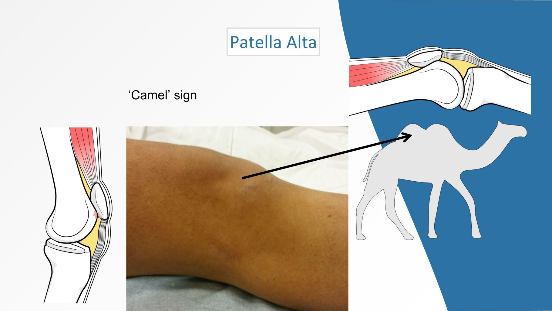
Camel knee – Camel hump knee.
If the kneecap is too high, it can often be characterized by the kneecap bulging up and then there is another bulge where the fat pad is located. The two bumps have earned it the name ‘camel knee’ as it resembles the camel’s two humps.
As mentioned above, too high a kneecap can cause anterior knee pain. You can read more about anterior knee pain here. Anterior knee pain is typically treated with physiotherapeutic exercise, but when the pain is caused by an overload of the fat body, the pain will often worsen and rest is a better solution.
Many people know that high-heeled shoes can leave marks on the floor because of the pressure on the heel. The same goes for cartilage, if there is too much pressure on a small area, the cartilage can’t take it.

This is the knee from the front. The yellow area illustrates the small area of overlap between the cartilage of the kneecap and the cartilage of the guide furrow. If there is less than 10 mm overlap, it can cause pain.
The cause of patella alta is unknown, but it is believed to be congenital.
Patient information leaflet tendon booklet relocation – no. 12
The treatment for a high kneecap
It is generally a very good idea to undergo surgery for patella alta if it causes symptoms. Patella alta is not a condition that can be corrected through exercise, such as physical therapy. You can reduce your activity level, which may help the pain subside. Patella alta predisposes the cartilage behind the kneecap and in the groove (trochlea) to wear and tear. This ultimately leads to osteoarthritis. Surgery can not only help stabilize the kneecap and reduce pain, but also prevent osteoarthritis.
A so-called dTTO cannot be performed while a child is still growing, as surgery risks damaging the upper growth plate in the tibia, which could lead to a deformity. Instead, the patellar tendon can be shortened in this age group. If the surgery cannot wait until the child has finished growing, this procedure is performed; however, it is not as safe as a dTTO, and without shortening the patellar tendon, it is a suboptimal procedure.
It sounds very simple: if the kneecap is positioned too high, it “just” needs to be moved downward. However, it requires a fair amount of knowledge and experience to avoid moving the kneecap too much or too little. If it is moved too far down, a condition called patella baja develops, which means the kneecap is positioned too low; this causes significant pain and prevents the knee from bending normally. Below, you can read about patellar tendon reattachment, which is a surgical procedure. This is currently probably the most common surgery I perform for anterior knee pain, and the results are really good,
Relocation of patellar tendon attachment because the kneecap is too high riding
If the kneecap is positioned too high relative to the groove (a condition called patella alta), this surgery can be used to lower the kneecap into the groove (a procedure called distalization of the tibial tuberosity). The surgeon can use an MRI scan to determine whether the overlap between the patellar cartilage and the trochlear cartilage is adequate (patellotrochlear index). If the patella is positioned too high, the overlap is too small (less than 6 mm).


Downward movement of the patellar tendon attachment is called distalization of the tibial tuberosity (dTTO). Most people ask “why don’t you just pull the tendon downwards”?
This is because it’s too demanding to get the tendon to heal into the bone. It is better to move the bone (tuberositas tibia), the place where the tendon is attached.
During surgery, the kneecap is brought down into the groove by pulling the patellar attachment further downwards and thus the kneecap also comes down. As you can see from the image on the left, the patellar tendon attachment is moved downwards and fixed with four titanium screws.
For many years, I have used 4 smaller screws instead of the standard 2 large screws.
The advantage is that the screw heads are significantly smaller and there is less risk of the screw heads bothering you when you kneel.
The 4-screw technique is stronger, allowing you to support the knee immediately. This helps you recover faster and reduces the risk of complications.
The 4-screw technique is so stable that it is not necessary to use a splint after surgery. The use of a splint can increase the risk of deep vein thrombosis.
Aim for an overlap between the cartilage of the kneecap and the cartilage of the furrow of approximately 50% when the knee is stretched.
The osteotomy is V-shaped, and the upper portion is performed with an osteotome (chisel) to prevent heat buildup, which overall results in faster and safer healing.
At the lower end, the osteotomy is performed with a saw to create a taper rather than a transverse cut, as this reduces the risk of the tibia fracturing.
Complications associated with dTTO may include tibial fractures and failure to heal. I have not observed these complications with this technique.
Using small, countersunk screws reduces the risk of pain over the screw heads, but it cannot be completely avoided. In rare cases, the screws must be removed because they cause pain. I have had to remove screws three times, which suggests that the risk is approximately 1%, given that I have performed several hundred of these surgeries.
Here is a link to Youtube concerning the discussion between Hoffa Impingingement and jumpers knee and patella alta – klik her
Complications of patellar tendon relocation
Unfortunately, not everyone recovers well after this type of surgery. If you’ve had surgery for a loose kneecap, some people unfortunately find that their kneecap remains loose. For those undergoing surgery for anterior knee pain, not everyone recovers completely, although the success rate is quite high for those who have their patellar tendon attachment moved downward due to a patella that is positioned too high. In a small percentage of cases, complications unfortunately occur. These can include pain and sensory disturbances over the operated patellar tendon attachment; in some cases, the bony block does not heal back together, and some patients develop an infection (however, I have never encountered the latter two complications, even though I have performed several hundred surgeries). Some patients experience sensory disturbances over the patellar tendon attachment site, and for some, the screws cause discomfort when they kneel—however, this is rare if four small-headed screws are used instead of two large-headed ones. Some patients ask if it is a complicated surgery, and it is not, provided the surgeon has extensive experience, uses the correct surgical technique, and possesses specialized knowledge in this particular area. The greatest risk arises if the surgeon uses outdated methods to assess the position of the patella, such as the Insall-Salvati ratio, the Caton-Dechamps ratio, or the Blackburn-Peel ratio. Using these methods carries the risk of diagnosing Patella Alta when it is not actually present. There is also a risk that the patellar attachments will be moved too far down during surgery, resulting in a condition called Patella Baja. In this condition, the patella sits too low, which can lead to pain and reduced knee flexion.
MRI Scan and Patella Alta
On an MRI scan, patella alta is best assessed by measuring the patellotrochlear index. This index requires correction for the degree of knee flexion seen in the scan images. Even though it may appear as if the knee is fully extended on an MRI scan, it is not actually fully extended. This is because during the MRI scan, a small device (called a coil) is placed behind the knee, causing the knee to bend by just a few—but crucial—degrees.
The reason why the overlap (patellotrochlear index) between the cartilage on the back of the patella and the groove (trochlea) is important is that it plays a key role in the biomechanics of the knee and the function of the cartilage.
Many people use the Caton-Dechamp or Insall-Salvati indices because they are the traditional measurement methods. These methods date back to a time when X-rays were the only diagnostic tool available. However, cartilage cannot be seen on X-rays, and therefore these measurement methods are outdated, even though they are still widely used. The Caton-Dechamp index is not entirely useless, however, as it has the advantage of not being affected by how much the knee is bent. On the other hand, it does not take into account the length of the trochlea.
<script type=”application/ld+json”>


